OBJECTIVES: The aim of the following study was to assess contour changes after socket preservation techniques.
MATERIAL AND METHODS: In five beagle dogs, the distal root of the third and fourth mandibular premolars was extracted. The following treatments (Tx) were randomly assigned for the extraction socket. Tx 1: BioOss Collagen. Tx 2: BioOss Collagen and a free soft tissue graft. Tx 3: No treatment. Tx 4: The internal buccal aspect was covered with an experimental collagen membrane, the extraction socket was filled with BioOss Collagen and the membrane folded on top of the graft. Impressions were obtained at baseline, 2 and 4 months after surgery. Bucco-lingual measurements were performed using digital imaging analysis.
RESULTS: All groups displayed contour shrinkage at the buccal aspect. Only the differences between the two test groups (Tx 1, Tx 2) and the control group (Tx 3) were significant at the buccal aspect (p< or =0.001). No measurements of the Tx 4 group could be performed.
CONCLUSION: Socket preservation techniques, used in the present experiment, were not able to entirely compensate for the alterations after tooth extraction. Yet, incorporation of BioOss Collagen seems to have the potential to limit but not avoid the post-operative contour shrinkage.
J Clin Periodontol. 2008 Oct;35(10):906-13. Epub 2008 Aug 17.
Showing posts with label ridge preservation. Show all posts
Showing posts with label ridge preservation. Show all posts

Ridge preservation techniques for implant therapy (Review JOMI)
PURPOSE:
The aim of this review was to evaluate the techniques and outcomes of postextraction ridge preservation and the efficacy of these procedures in relation to subsequent implant placement.MATERIALS AND METHODS:
A MEDLINE/PubMed search was conducted and the bibliographies of reviews from 1999 to March 2008 were assessed for appropriate studies. Randomized clinical trials, controlled clinical trials, and prospective/retrospective studies with a minimum of five patients were included.RESULTS:
A total of 135 abstracts were identified, from which 53 full-text articles were further examined, leading to 37 human studies that fulfilled the search criteria. Many different techniques, methodologies, durations, and materials were presented in the publications reviewed, making direct comparison difficult.CONCLUSIONS:
Despite the heterogeneity of the studies, it was concluded that ridge preservation procedures are effective in limiting horizontal and vertical ridge alterations in postextraction sites. There is no evidence to support the superiority of one technique over another. There is also no conclusive evidence that ridge preservation procedures improve the ability to place implants.

Int J Oral Maxillofac Implants. 2009;24 Suppl:260-71.
Ridge preservation techniques for implant therapy.
Socket Augmentation: Rationale and Technique (Part 3)
Guided Bone Regeneration (GBR)
Cases in which the buccal plate is absent or was lostduring exodontia require a different approach.GBR techniques associated with or without immediate implant placement are required to treat these ridge
defects. A delayed implant placement approach is indicated when primary stability of the implant cannot be predictably achieved, particularly in sockets of bi- or multirooted teeth.
In these cases GBR techniques, such as the “sandwich bone augmentation,” are indicated (Wang et al. 2004b).
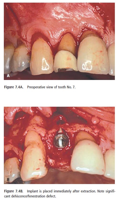

Abd El Salam El Askary, Fundamentals of Esthetic Implant Dentistry, 2007
Socket Augmentation: Rationale and Technique (Part 2)
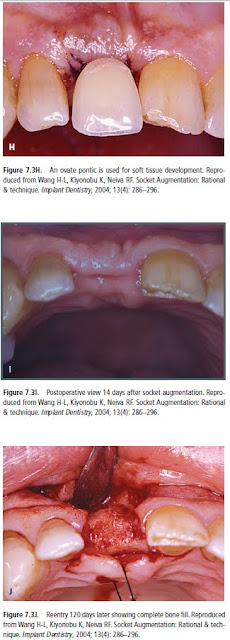
Layers Technique (when buccal plate is ≤1 mm thickness)
The layers technique was developed to maximize bone healing in sockets with compromised healing potential.
A combination of a bone replacementgraft and a collagen wound dressing material should be used when the buccal socket wall is ≤1 mm thick at approximately 2–3mm below the alveolar crest and/or occurrence of dehiscence or fenestrations are found. Mineralized bone grafts that are quickly replaced by host bone are
preferable.
The bone graft should be tamped down lightly, and overfill should be avoided. Adequate space between the graft particles is critical to allow for revascularization to spread throughout the graft, bringing the proteins and growth factors necessary for new bone growth (Becker et al. 1992, Mellonig 1996).
The histologic data obtained from our unpublished data indicated 68% of vital bone, 5% of residual particle, and 27% of connective tissue. This is similar to the human host bone component. Sites that may also benefit from using this technique include those with thin bone ≤1 mm, periapical pathologies, and sockets of multirooted teeth associated with loss of the interradicular bone.


The histologic data obtained from our unpublished data indicated 68% of vital bone, 5% of residual particle, and 27% of connective tissue. This is similar to the human host bone component. Sites that may also benefit from using this technique include those with thin bone ≤1 mm, periapical pathologies, and sockets of multirooted teeth associated with loss of the interradicular bone.
A combination of a bone replacementgraft and a collagen wound dressing material should be used when the buccal socket wall is ≤1 mm thick at approximately 2–3mm below the alveolar crest and/or occurrence of dehiscence or fenestrations are found. Mineralized bone grafts that are quickly replaced by host bone are
preferable.
The bone graft should be tamped down lightly, and overfill should be avoided. Adequate space between the graft particles is critical to allow for revascularization to spread throughout the graft, bringing the proteins and growth factors necessary for new bone growth (Becker et al. 1992, Mellonig 1996).
The histologic data obtained from our unpublished data indicated 68% of vital bone, 5% of residual particle, and 27% of connective tissue. This is similar to the human host bone component. Sites that may also benefit from using this technique include those with thin bone ≤1 mm, periapical pathologies, and sockets of multirooted teeth associated with loss of the interradicular bone.


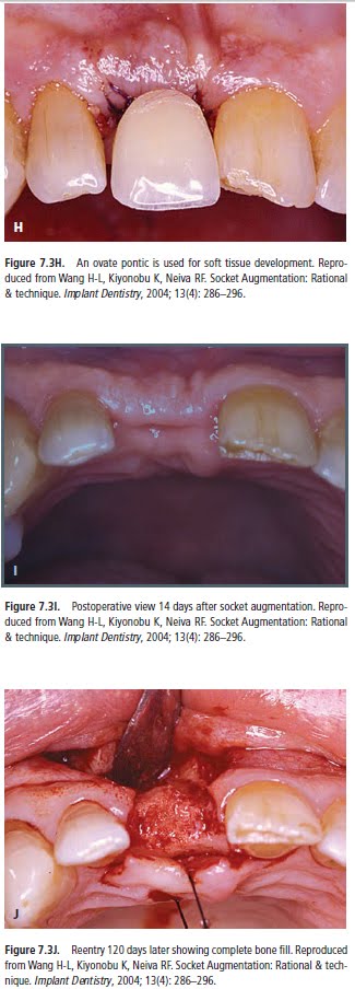
The histologic data obtained from our unpublished data indicated 68% of vital bone, 5% of residual particle, and 27% of connective tissue. This is similar to the human host bone component. Sites that may also benefit from using this technique include those with thin bone ≤1 mm, periapical pathologies, and sockets of multirooted teeth associated with loss of the interradicular bone.
Abd El Salam El Askary, Fundamentals of Esthetic Implant Dentistry, 2007
Future posts will describe cases, in which the buccal plate is absent or was lost during exodontia require a different approach
Socket Augmentation: Rationale and Technique
Tooth extraction, either traumatic or atraumatic, results in alveolar bone loss, both in width and height (Bays
1986, Mecall and Rosenfeld 1996, Mecall and Rosenfeld 1992, Mecall and Rosenfeld 1991). An average of 40% to 60% of original height and width is expected to be lost after tooth extraction, with the greatest loss happening within the first year (Sevor and Meffert 1992, Polizzi et al. 2000, Grunder et al. 1999, Werbitt and Goldberg 1992, Werbitt and Goldberg 1991). This can negatively influence bone volume that is needed for future dental implant placement as well as proper ideal esthetic restoration. Research has demonstrated that the alveolar ridge at the maxillary anterior area can be reduced by 23% in the first six months after tooth extraction, and an additional 11% in the following five years (Artzi et al. 2000). In the posterior mandible, resorption happens primarily in the buccal/labial direction, resulting in a lingual displacement of alveolar crest (Artzi et al. 2000).
Traditional Socket Management with or without Collagen Wound Dressing Material (socket management when
buccal plate is ≥1 mm thickness):


1986, Mecall and Rosenfeld 1996, Mecall and Rosenfeld 1992, Mecall and Rosenfeld 1991). An average of 40% to 60% of original height and width is expected to be lost after tooth extraction, with the greatest loss happening within the first year (Sevor and Meffert 1992, Polizzi et al. 2000, Grunder et al. 1999, Werbitt and Goldberg 1992, Werbitt and Goldberg 1991). This can negatively influence bone volume that is needed for future dental implant placement as well as proper ideal esthetic restoration. Research has demonstrated that the alveolar ridge at the maxillary anterior area can be reduced by 23% in the first six months after tooth extraction, and an additional 11% in the following five years (Artzi et al. 2000). In the posterior mandible, resorption happens primarily in the buccal/labial direction, resulting in a lingual displacement of alveolar crest (Artzi et al. 2000).
Traditional Socket Management with or without Collagen Wound Dressing Material (socket management when
buccal plate is ≥1 mm thickness):


Abd El Salam El Askary, Fundamentals of Esthetic Implant Dentistry, 2007
Future posts will describe cases, when buccal plate is ≤1 mm thickness and Cases in which the buccal plate is absent or was lost during exodontia require a different approach
Subscribe to:
Posts (Atom)