Layers Technique (when buccal plate is ≤1 mm thickness)
The layers technique was developed to maximize bone healing in sockets with compromised healing potential.
A combination of a bone replacementgraft and a collagen wound dressing material should be used when the buccal socket wall is ≤1 mm thick at approximately 2–3mm below the alveolar crest and/or occurrence of dehiscence or fenestrations are found. Mineralized bone grafts that are quickly replaced by host bone are
preferable.
The bone graft should be tamped down lightly, and overfill should be avoided. Adequate space between the graft particles is critical to allow for revascularization to spread throughout the graft, bringing the proteins and growth factors necessary for new bone growth (Becker et al. 1992, Mellonig 1996).
The histologic data obtained from our unpublished data indicated 68% of vital bone, 5% of residual particle, and 27% of connective tissue. This is similar to the human host bone component. Sites that may also benefit from using this technique include those with thin bone ≤1 mm, periapical pathologies, and sockets of multirooted teeth associated with loss of the interradicular bone.


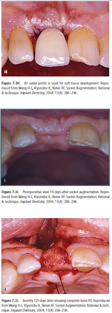
The histologic data obtained from our unpublished data indicated 68% of vital bone, 5% of residual particle, and 27% of connective tissue. This is similar to the human host bone component. Sites that may also benefit from using this technique include those with thin bone ≤1 mm, periapical pathologies, and sockets of multirooted teeth associated with loss of the interradicular bone.
A combination of a bone replacementgraft and a collagen wound dressing material should be used when the buccal socket wall is ≤1 mm thick at approximately 2–3mm below the alveolar crest and/or occurrence of dehiscence or fenestrations are found. Mineralized bone grafts that are quickly replaced by host bone are
preferable.
The bone graft should be tamped down lightly, and overfill should be avoided. Adequate space between the graft particles is critical to allow for revascularization to spread throughout the graft, bringing the proteins and growth factors necessary for new bone growth (Becker et al. 1992, Mellonig 1996).
The histologic data obtained from our unpublished data indicated 68% of vital bone, 5% of residual particle, and 27% of connective tissue. This is similar to the human host bone component. Sites that may also benefit from using this technique include those with thin bone ≤1 mm, periapical pathologies, and sockets of multirooted teeth associated with loss of the interradicular bone.


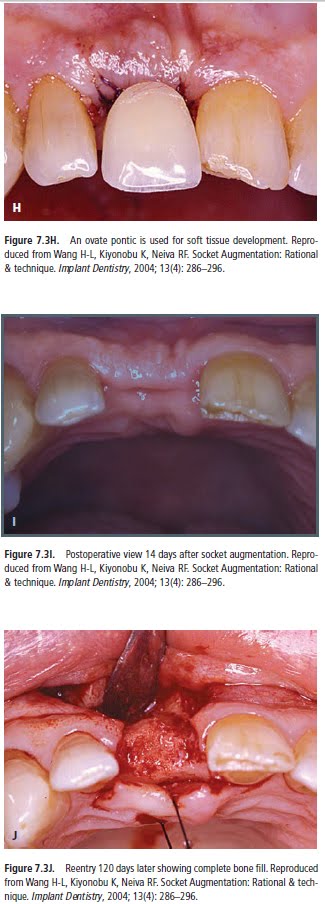
The histologic data obtained from our unpublished data indicated 68% of vital bone, 5% of residual particle, and 27% of connective tissue. This is similar to the human host bone component. Sites that may also benefit from using this technique include those with thin bone ≤1 mm, periapical pathologies, and sockets of multirooted teeth associated with loss of the interradicular bone.
Abd El Salam El Askary, Fundamentals of Esthetic Implant Dentistry, 2007
Future posts will describe cases, in which the buccal plate is absent or was lost during exodontia require a different approach


